


The term osteochondrosis itself is derived from two words: osteo - bone and chondrue - cartilage. Simply put, it is the process of cartilage. Although this interpretation is fundamentally wrong. Some people go even further, believing that osteonecrosis is the deposition of salt in the joints. Furthermore, it is table salt that is said to be consumed in large quantities for food.
Pathogenesis
In reality, things turn out a little differently. And more difficult. And table salt, if it plays any role in the occurrence of osteonecrosis, it's very indirect. Osteoarthritis is based on the degeneration and degeneration of articular cartilage. This is not an independent disease, but a pathological process that can be noted almost anywhere where connective cartilage tissue is present.
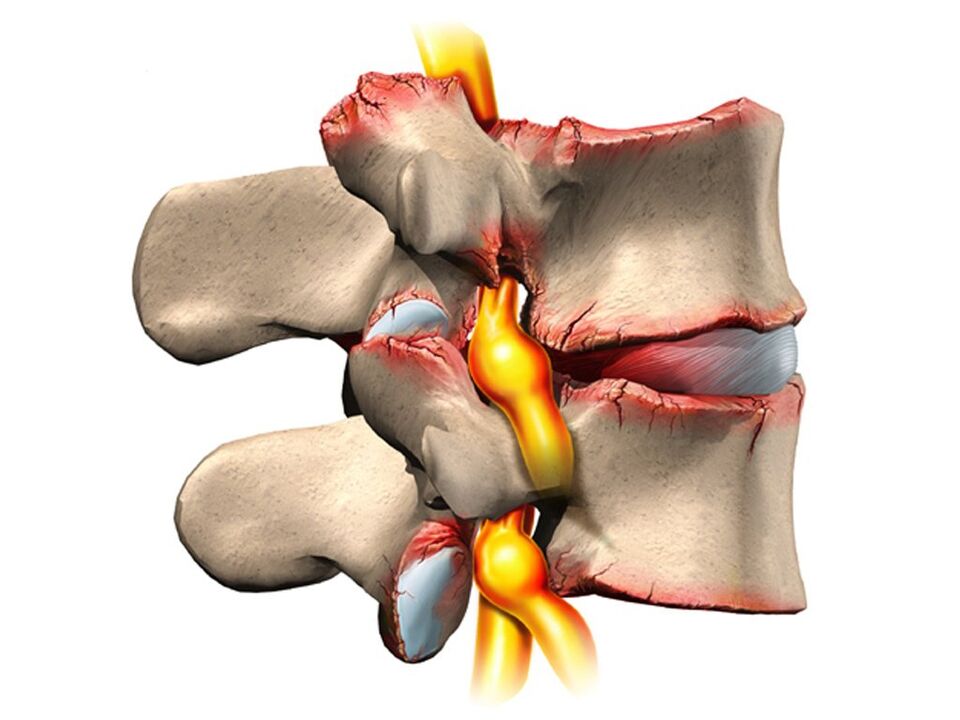
However, osteonecrosis in overwhelming cases affects the spine. Why so? The fact is that between the vertebrae there is a type of cushion - intervertebral discs (discs). The physiological role of these discs is to cushion and protect the vertebral bodies from premature wear from mechanical impact. The intervertebral disc consists of a loose inner nucleus surrounded by an arcuate fiber and an upper and lower cushion.
The disc is subjected to tremendous mechanical stress, resulting in permanent damage to its structures at the cellular level. In humans, these processes are all too obvious - this is our payment for vertical walking. To prevent the disk from being "erased" completely, it must constantly regenerate, i. e. rebuild itself. It is the balance of injury-regenerating processes that determines the normal structure of the disc. Another curious detail is that the supply of blood and nutrients to the intervertebral discs is not accomplished through the overgrown blood vessels in childhood but diffuses from the bony tissue of the vertebral bodies. Again, the payout for mobility is with two limbs, not four.
Therefore, the intervertebral discs are anatomically and physiologically vulnerable. Any negative process in the body leads to an imbalance in the balance of injury regeneration, and leads to the development of dystrophy and degeneration in the intervertebral disc. A structurally defective disc can no longer withstand proper mechanical stress. Under extreme pressure from the upper vertebrae, the discs are displaced in different directions, usually laterally and posteriorly. This process is called a herniated disc.
The bony tissue of the vertebrae that has lost its cartilage lining is also subjected to mechanical wear and tear. Due to constant trauma to the anterior edge surface of the vertebral bodies, pathological foci are formed - osteomas. Spondylolisthesis develops. Due to the degeneration and displacement of the discs, the disc space is reduced, the spinal canal narrows and the roots of the spinal nerves in the so-called foramina foramina are compromised.
Reason
The causes, or etiological factors, of osteonecrosis are diverse. They can be both local, that is, due to pathology of the spine itself and general disorders at the organ level. Any pathology that leads to a violation of the structure of the spine or a metabolic disorder can be considered the cause of osteonecrosis. In this regard, there are:
- Changes in the configuration of the spine (scoliosis, scoliosis, or scoliosis).
- Other malformations of the musculoskeletal system are flat feet, narrow shoulders, and abnormal pelvic structures.
- Spinal cord injury.
- Weak immunity.
- Metabolic disorders - osteoporosis, obesity, diabetes mellitus, thyroid disease.
- Diseases of the cardiovascular system - atherosclerosis, hypertension.
- Digestive disorders lead to inadequate absorption of nutrients from the gastrointestinal tract.
- Heredity.
It should be noted that these conditions do not necessarily lead to osteonecrosis. This requires constant exposure to several risk factors - hypothermia, malnutrition, a sedentary lifestyle, or conversely, excessive physical exertion.
Symptom
Osteochondrosis is itself an asymptomatic process. At the same time, signs of disc degeneration are also very diverse. How to so? The fact is that the clinical manifestations of osteonecrosis are based on its complications - herniated disc, spondylolisthesis, sciatica, spinal stenosis.
Furthermore, the clinic is highly variable depending on the predominant location of the process in the cervical, thoracic or dorsal spine. The last part is most often affected, as the lower back is where physical activity is maximal. Signs of light zone osteonecrosis:
- Pain (paralysis, low back pain, sciatica).
- Limit movement of the lower back and lower extremities (intermittent movements).
- Here, sensitization disorders of the paresthesia type - numbness, burning, gauze.
- Pathological lumbar muscle strain.
- In the absence of treatment, dysfunction of the pelvic organs.
Cervical fibroids are observed less often than cervical fibroids. However, this disease is also quite common. In addition to typical signs such as pain (cervical pain), decreased sensitivity and movement in the upper extremities, tarsal osteosarcoma caused by impaired blood supply to the brain has its own characteristics. These features are demonstrated:
- Insomnia.
- Headache, dizziness.
- Periodic nausea.
- General weakness, rapid fatigue.
- Blood pressure fluctuations.
- Occasional toothache.
- Behavioral reactions in the form of tears, irritability.
The affected area of the chest with osteonecrosis is relatively rare. The patients in this case are people who are forced by their profession to sit in an uncomfortable fixed position - schoolchildren, students, programmers, office workers. Symptoms of osteonecrosis in this case will be as follows:
- Pain and paresthesia in the chest.
- Shortness of breath.
- Heartbeat feeling.
- Limit movement of the thoracic spine.
Diagnose
From all this, it is clear that osteonecrosis is a chameleon disease. Because of the same signs, it is easy to confuse with cerebrovascular accident, hypertension, myocardial infarction, angina pectoris, autonomic nervous disorder. That is why, for an accurate diagnosis, a comprehensive and complex diagnosis is necessary to accurately identify the symptoms and treat the osteonecrosis.
This diagnosis, in addition to traditional questioning and clarifying patient complaints, should include a medical examination and special research methods. These methods include X-ray of the spine, ultrasound of internal organs. Recently, computer imaging and magnetic resonance have been successfully used to diagnose osteonecrosis.
Treatment
Therapeutic strategies for osteonecrosis include the use of:
- Medicines.
- Massage.
- Physiotherapy procedures.
- Physical therapy (exercise therapy).
- Manual therapy.
- Acupuncture.
Drugs for the treatment of osteonecrosis are mainly aimed at reducing pain and eliminating inflammatory processes in the nerve roots. In various combinations, these drugs are widely used in the form of ointments, injections, and tablets to treat osteonecrosis. It should not be forgotten that these drugs have an adverse effect on the liver, stomach and intestines. In this way, they can exacerbate the metabolic disorders in osteonecrosis. They relieve pain due to good blockade with local anesthetics. Yes, the effects of these funds are short-lived, and have no effect on the overall process of osteonecrosis.
It is possible to improve metabolism at the body and local level with the help of drugs such as chondroprotectors, immunostimulants and vitamins with minerals. Chondroprotectors are used in tablets, ointments and ampoules. Among the fortifiers, vitamin C, group B, in combination with minerals are used. In this regard, calcium preparations are preferred above all. Indeed, contrary to some misconceptions, the basis of osteonecrosis is not an excess, but only a deficiency of calcium.
After successful exacerbation, physiotherapeutic procedures, massage, and exercise are shown. As physical procedures, calcium electrophoresis, hydrocortisone electrophoresis, amplipulse, paraffin therapy are used. All these measures are aimed at eliminating pain and inflammation in nerve roots, ligaments and muscles. Massage therapy for osteonecrosis is performed according to a generally accepted method. The massage area is selected depending on the location of the osteonecrosis. The extension of range of motion is achieved with the help of exercise therapy. At the beginning, during the exacerbation phase, there is practically no dynamic load. The patient is constantly in an optimal position. At this time, it is desirable to wear immobilizers - a corset, a Shants' collar. As the exacerbation subsides, the volume and duration of movements in exercise therapy increase.
Recently, in the treatment of osteonecrosis, non-traditional methods of treatment have been applied - acupuncture, manual therapy, osteopathy. Acupuncture is the impact on special biologically active points located along the spine, on the backs of hands and feet. With manual therapy, the normal position of the vertebrae and discs is restored through the manual action of the therapist's hand. And during osteopathy, the structural integrity of the musculoskeletal system is ensured using specific techniques. In the absence of effect of conservative measures to treat osteonecrosis, persistent pain, complications, surgery is indicated. Pathologically displaced discs are removed. Currently, for this purpose, microdiscectomy is performed - endoscopic removal of a displaced disc.